Figure 5.

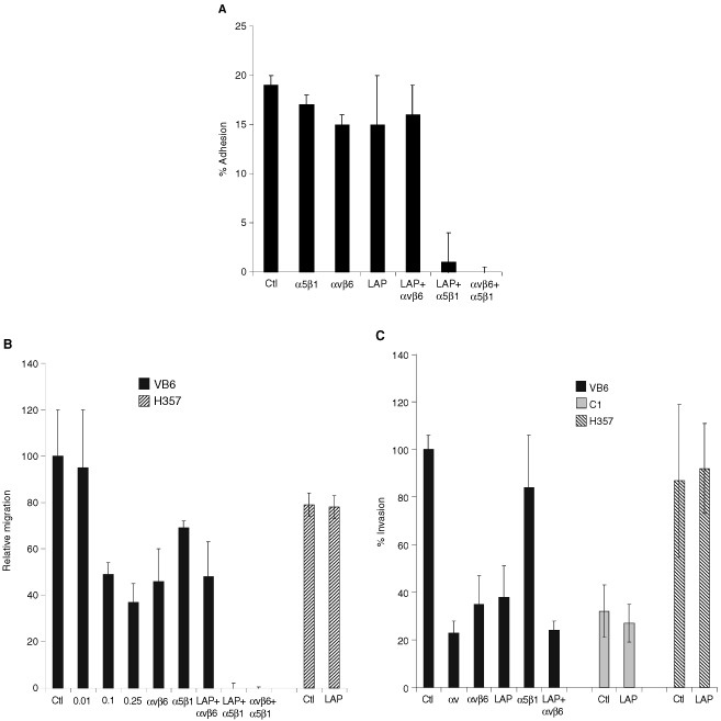
Soluble LAP inhibits αvβ6 interaction with fibronectin. (A) LAP inhibits VB6 adhesion to fibronectin. Chromium [51Cr]-labelled VB6 cells were added to fibronectin-coated 96-well plates containing an irrelevant control antibody (W632) or test antibodies against αvβ6 (10D5), α5β1 (P1D6) and LAP (0.25 μg ml−1) or combinations thereof. Figure shows a representative experiment performed in quadruplicate. Results are expressed relative to binding in the presence of a control antibody (=100). Background binding to BSA has been subtracted from the results. Error bars represent standard deviation. VB6 cells adhere to fibronectin through αvβ6 and α5β1 and a combination of antibodies against both integrins is required to block adhesion completely (Thomas et al, 2001b). Treating the cells solely with LAP, or antibodies against αvβ6 or α5β1 did not inhibit adhesion significantly (neither did a combination of LAP and anti-αvβ6). However, combinations of anti-α5β1 and LAP or anti-αvβ6 and anti-α5β1 inhibited adhesion completely. These data confirm that soluble LAP produces a similar effect to anti-αvβ6 antibody and suggests that LAP may alter keratinocyte binding to fibronectin through its interaction with the αvβ6 integrin. (B) LAP inhibits VB6 migration to fibronectin. To determine the effect of LAP on cell migration, the cells were treated with varying concentrations of LAP peptide prior to plating and then allowed to migrate for 8 h before counting. A representative experiment performed in quadruplicate is shown. Results are expressed relative to migration in the presence of a control antibody (=100). Error bars represent standard deviation. Haptotactic migration of VB6 cells towards fibronectin is modulated through αvβ6 and α5β1 integrins and maximal inhibition of migration is produced by blocking both integrins (Thomas et al, 2001b). Migration of VB6 cells towards fibronectin was inhibited by a concentration of LAP as low as 0.1 μg ml−1 (51% inhibition). At higher concentrations a further degree of inhibition was seen (0.25 μg ml−1=63% inhibition). This level of inhibition was similar to that produced by anti-αvβ6 antibody (54% inhibition) and did not differ significantly from a combination of LAP and anti-αvβ6 antibody (52% inhibition). Complete inhibition of migration was produced using a combination of soluble LAP with anti-α5β1 antibody (or a combination of anti-αvβ6 with anti-α5β1 antibody). Soluble LAP had no effect of the migration of H357 αv-negative cells. These data confirm that soluble LAP produces a similar effect to anti-αvβ6 antibody and suggest that LAP may inhibit αvβ6-dependent keratinocyte migration towards fibronectin. (C) LAP inhibits αvβ6-dependent invasion through Matrigel. Cell invasion assays were performed over 72 h using Matrigel coated polycarbonate filters. To assess the effect of soluble LAP invasion, cells were treated with LAP (0.5 μg ml−1) for 30 min at 4°C prior to plating. Following incubation the cells in the lower chamber (including those attached to the undersurface of the membrane) were trypsinised and counted on a Casy 1 counter (Sharfe System GmbH, Germany). Figure shows a representative experiment performed in quadruplicate. Error bars represent standard deviation. Pre-incubation of VB6 cells with soluble LAP inhibited invasion (62% inhibition), producing a similar level of inhibition to anti-αvβ6 antibody (58% inhibition). Soluble LAP had no effect on invasion of H357 αv-negative cells or C1 control cells which express low levels of endogenous αvβ6. These data suggest that soluble LAP may inhibit αvβ6-dependent cell invasion.
