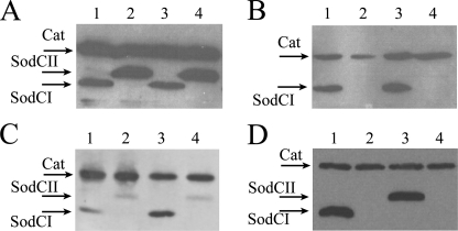
FIGURE 5.
In vitro and in vivo SodCI and SodCII accumulation. Bacterial lysates were loaded on a 12% polyacrylamide gel, which was processed for Western blot analysis. Membranes were probed with anti-FLAG monoclonal antibodies. Strains MA7224 (lane 1), MA7225 (lane 2), SA167 (lanes 3), and SA168 (lane 4) were grown overnight in LB medium (panel A) or collected from J774.1 macrophages 23 h post-infection (panel B). Panel C, strains MA7224 (lanes 1 and 3) and MA7225 (lanes 2 and 4) were harvested from Caco-2 or THP-1 cells 23 h post-infection. Panel D, intracellular accumulation of SodCI and SodCII expressed from MA7224 (lane 1), MA7225 (lane 2) MA7537 (lane 3), and MA7538 (lane 4) in bacteria harvested from J774.1 macrophages 23 h post-infection. Cat, chlorampheniol acetyl transferase.

