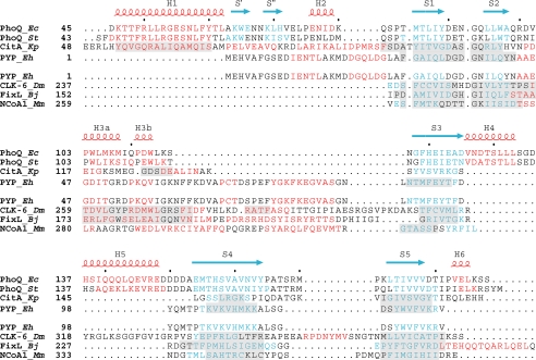
FIGURE 3.
Structure-based sequence alignments. Structure-based sequence alignments comparing PDC to PAS domains. Residues in helices are colored red, and residues in strands are colored blue. Secondary structure elements of E. coli PhoQ are also shown and labeled above the alignments, with helices in red and strands in blue.Inthe top set, the PDC sensor domains PhoQ and CitA are aligned with the PYP PAS domain. Regions in CitA and PYP having Cα positions structurally aligned with those in E. coli PhoQ are shaded in light gray.Inthe bottom set, the PAS domains CLK-6, FixL, and NcoA-1 are aligned with PYP. Regions in CLK-6, FixL, and NcoA-1 having Cα positions structurally aligned with those of PYP are shaded in light gray. Organism names are abbreviated in italics (Ec for E. coli, St for S. typhimurium, Kp for Klebsiella pneumoniae, Eh for Ectothiorhodospira halophila, Dm for Drosophila melanogaster, Bj for Bradyrhizobium japonicum, and Mm for Mus musculus).

