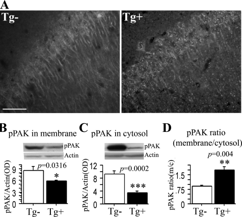
FIGURE 2.
PAK translocation in Tg2576 mice. A, immunofluorescent staining of PAK translocation in hippocampus of Tg2576 mice. 22-Month-old Tg2576 transgenic Tg+ and control Tg- mice were stained with anti-pPAK antibody. Anti-pPAK identified a loss of diffuse perikaryal and dendritic labeling along with granular structures in Tg2576 transgenic Tg+ mice; scale bar, 25 μm. B-D, Western immunoblot analysis of PAK translocation in the cortex in aged Tg2576 mice. The levels of pPAK were significantly reduced in membrane and cytosol from Tg2576 Tg+ mice when compared with control Tg2576 Tg- mice (B,*, p < 0.05; C, ***; p < 0.001), and in contrast, the pPAK ratio of membrane to cytosol was significantly increased in Tg2576 Tg+ mice when compared with control Tg2576 Tg- mice (D, ***, p < 0.001).

