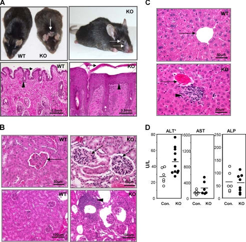
FIGURE 2.
Adult Mta2 knock-out (KO) mice exhibit autoimmunity-related phenotypes in multiple organs. A, upper panels, Mta2 KO mice show skin lesions at eyelid, mouth, nose, and neck area as indicated by arrows; lower panels, hemotoxylene and eosin (H&E) staining of skin sections from wild-type and Mta2 null mice show significant hyperkerotosis (arrow) and epidermal cell hyperplasia (acanthosis) (arrowheads)in Mta2 null mice. B, upper panels, H&E-stained kidney sections of Mta2 null mice reveal mesangial cell proliferation (arrowhead); lower panels, Mta2 null mice also show lymphocyte infiltration (arrow) in kidney. C, Mta2 null mice develop liver inflammation. H&E-stained liver sections of Mta2 null mice reveal lymphocyte infiltration (lower panel, arrowhead). Portal veins are indicated by arrows. D, comparison of the liver enzyme activity levels in serum measured by ELISA. Mta2 null mice had a significantly increased alanine aminotransferase (ALT) level (p < 0.01), indicating liver cell damage in these mice. The mutant mice showed no significant change in the levels of aspartate aminotransferase (AST) and alkaline phosphatase (ALP) in the serum of Mta2 null mice when compared with the wild-type mice. Asterisk indicates the difference is statistically significant.

