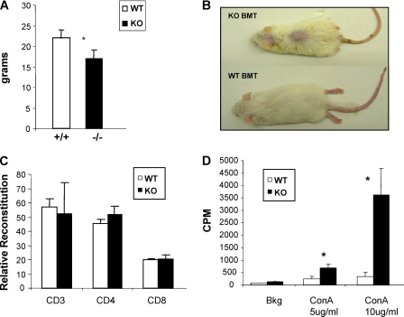
FIGURE 4.
Mta2-deficient BM cells cause decreased bodyweight, skin lesion, and T cell hyperproliferation in recipient SCID mice. A, the transfer of BM cells from Mta2 knock-out (KO) mice cause 25% decreased bodyweight in recipient SCID mice 8 weeks after bone marrow transplantation (p < 0.03). B, a representative SCID mouse received Mta2 KO BMT showed skin lesion (upper mouse), whereas the representative SCID mouse received BMT from wild-type mice exhibited normal skin (lower mouse) 8 weeks after transplantation. In this experiment, all three SCID mice that received Mta2 KO BM cells developed skin lesion and none of the four SCID mice received wild-type BM cells developed skin lesion. C, FACS analysis indicated that transplanted Mta2 KO BM cells successfully reconstituted in T cells in SCID mice. The relative reconstitution of T cells from Mta2 KO BMT SCID mice and WT BMT SCID mice were plotted (n = 3 for each genotype). D, the LN cells from Mta2 KO BMT SCID mice showed hyperproliferation upon different concentrations of concanavalin A (ConA) stimulation. Asterisk indicates the difference is statistically significant (p < 0.05).

