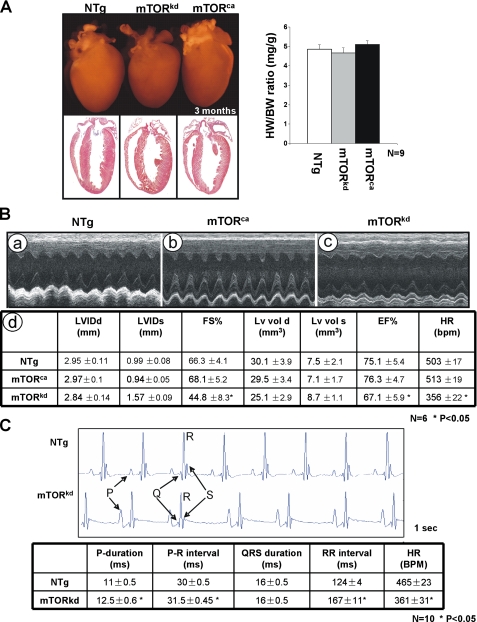
FIGURE 2.
Characterization of αMHC-mTORkd and αMHC-mTORkd transgenic mice. A, left panel, comparison of the gross morphology and histology of αMHC-mTORkd and αMHC-mTORca transgenic and littermate nontransgenic (NTg) control heart. There was no obvious morphological defect in both αMHC-mTORkd and αMHC-mTORca transgenic hearts when compared with littermate controls. Right panel, quantitative comparison of the ratio of heart weight versus body weight of αMHC-mTORkd and αMHC-mTORca transgenic hearts and nontransgenic littermate control. B, echocardiograph analysis of αMHC-mTORkd and αMHC-mTORca transgenic mice and littermate controls (3-month-old male). Representative M-Mode images are shown in panels a–c. Measurements of various parameters and statistics analysis are summarized in panel d. C, ECG recording of the αMHC-mTORkd and littermate control mice (3-month-old male). There was a difference in the function of atria and sinoatrial node in αMHC-mTORkd hearts compared with littermate controls.

