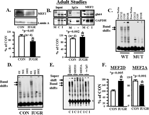
FIGURE 3.
Adult studies, MEF2 nuclear protein and glut4 DNA. A, top panel, representative Western blot demonstrating total nuclear MEF2 protein concentrations in CON and IUGR skeletal muscle with the nuclear marker Lamin A protein serving as an internal loading control. Bottom panel, quantification of MEF2 protein concentrations as a ratio to Lamin A protein is depicted as a percent of CON. Inter-group difference was assessed by Student's t test (*). B, top panel, representative 2% agarose gels demonstrate the input PCR glut4 and GAPDH control without an antibody, in the presence of nonspecific (–) and anti-polymerase (+) IgGs and ChIP assay demonstrating the 384-bp PCR glut4 and the 230-bp PCR GAPDH DNA amplification products obtained from total MEF2 nuclear chromatin IPs. M = DNA size markers, C = control, and I = IUGR. Bottom panel, quantification of the ChIP glut4 amplification product represented as a ratio to that of GAPDH, corrected for the input control, and shown as a percent of CON. Inter-group difference is assessed by Student's t test (*). C, representative polyacrylamide gel demonstrating two gel-shifted bands (arrows) in the presence of a 32P-end-labeled DNA probe (free probe) that spans the glut4 gene containing the wild type (WT) MEF2-binding site that are not seen in the presence of MUT MEF2-binding site in C2C12 cell (positive control), CON and IUGR skeletal muscle nuclear extracts. D, representative polyacrylamide gel demonstrating competition for the two gel-shifted bands (arrows) in the presence of a 32P-end-labeled DNA probe that spans the glut4 gene containing the wild type MEF2-binding site and skeletal muscle nuclear extracts from CON and IUGR groups with increasing concentrations (10× to 100×) of unlabeled probe. E, representative polyacrylamide gel demonstrating supershifted bands (double arrow) in the presence of a 32P-end-labeled glut4 DNA probe containing the wild type MEF2-binding site, skeletal muscle nuclear extracts from control (C) and IUGR (I) groups, and anti-MEF2C, anti-MEF2D, anti-MEF2A IgGs and in the absence of IgG (–ve). F, quantification of the MEF2D and MEF2A supershift bands in control (CON) and IUGR groups. Difference between the two groups was assessed by Student's t test (*).

