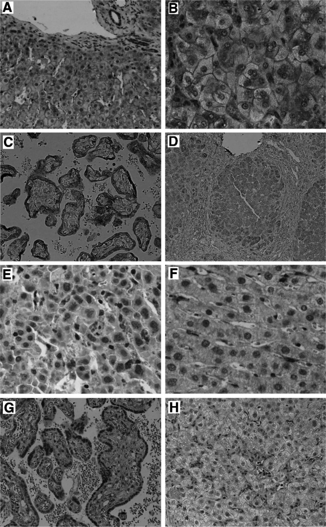
Figure 3.

Immunostaining for the insulin receptor on normal (A) and cirrhotic liver (B) with monoclonal antibody CT-3, producing a membraneous staining without obvious differences between architecturally normal (A) and cirrhotic (B) liver. Immunostaining for the IGF-1R revealed a trophoblast epithelial staining pattern in mature placenta (C), and weak cytoplasmic and membraneous staining largely of hepatocytes in normal and cirrhotic liver (D). The IGFBP-3-specific monoclonal antibody reacted with sinusoidal cells on cirrhotic (E) and normal liver (F), part of which is morphologically compatible with Kupffer cells. Immunostaining for the IGFBP-4 on placenta for control (G) and cirrhotic liver (H) with the anti-human IGFBP-4 antibody. The IGFBP-4-specific antibody showed a clear cytoplasmatic staining in mature placenta (G), whereas cirrhotic liver displayed a weak immunoreactivity of cells also compatible with Kupffer cells.
