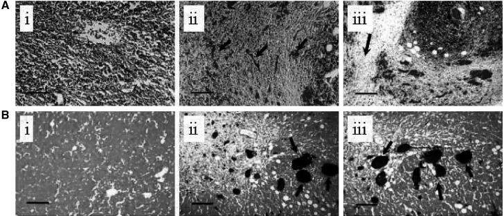
Figure 2.
Histological features of spontaneous regression in E.G7-OVA tumours. Tumour sections (5 μm thick) were stained with Masson's trichrome (A) or carmine red dye counterstained with light green (B). In A (ii and iii) the tumour cells can be seen to be surrounded by fibroblasts and capillaries filled with red blood cells (arrowed). There is also evidence of collagen deposition, which stains green with this dye (arrowed in A iii). A(i) is a representative section from an EL-4 tumour. The tumour sections shown in B (ii and iii) were obtained following injection of the animal with carmine dye. Functional vessels are stained red in these sections (arrowed). B(i) is a representative section from an EL-4 tumour. The bars are 400 μm long.

