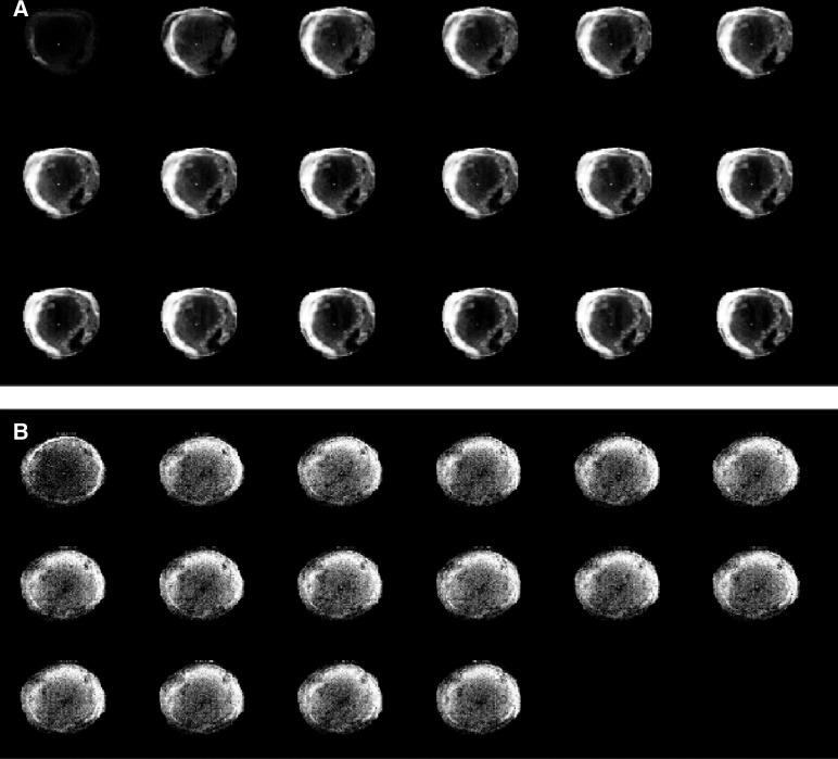
Figure 3.
A series of T1-weighted MR images acquired from a regressive E.G7-OVA tumour (A) and an EL-4 tumour (B) following i.v. injection of the MRI contrast agent, Gd-DTPA. The first images in the series were acquired prior to contrast agent injection. The subsequent images (reading from left-to-right and top-to-bottom) were acquired at 2 min intervals. The presence of the contrast agent increases signal intensity in the images and these increases are proportional to its concentration.

