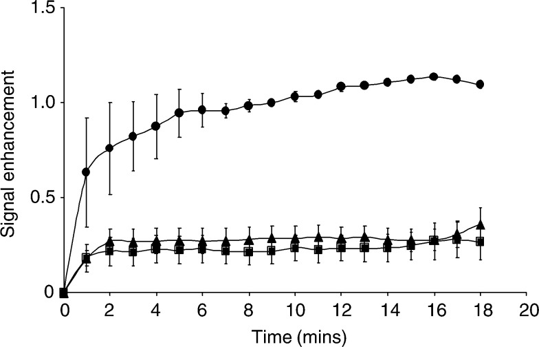
Figure 5.
Time-dependent changes in signal enhancement in tumours using dynamic contrast-enhanced MRI with the macromolecular contrast BSA–Gd-DTPA. A series of T1-weighted spin echo images were acquired from EL-4 (▴, n=7), progressive E.G7-OVA (▪, n=6) and regressive E.G7-OVA (•, n=2) tumours. The symbols and error bars show the mean±s.e.m. The values were calculated for a 20 pixel-wide band in the tumour peripheries, where vessel density was higher. The high molecular weight agent is initially confined to the vasculature, so the greater signal enhancement of regressive E.G7-OVA tumours is indicative of a higher functional vascular volume fraction.

