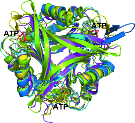
Figure 5.
Ribbon diagram of the trimer of HsCutA1 (coloured in green) overlaid with PII proteins of similar structure. The PII proteins are from Methanococcus jannaschii (PDB code 2j9d; Yildiz et al., 2007 ▶; marine), Synechococcus elongates (2v5h; Llácer et al., 1991 ▶; yellow), Bacillus cereus (2gx8; Godsey et al., 2007 ▶; magenta) and E. coli (2gnk; Xu et al., 1998 ▶; cyan). The molecules of ATP bound to E. coli GlnK (2gnk; Xu et al., 1998 ▶) in the interface clefts are presented as ball-and-stick models.

