
Figure 1.
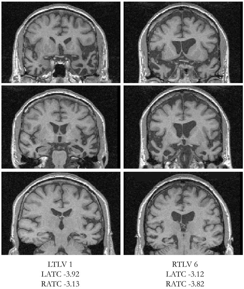
Coronal T1 MP-RAGE images of a representative pairing of patients with left and right temporal lobe variants (LTLV and RTLV) using temporal volume Z scores. The left side of each image represents the right side of the head. LATC = left anterior temporal cortex; RATC = right anterior temporal cortex.