
Figure 1.
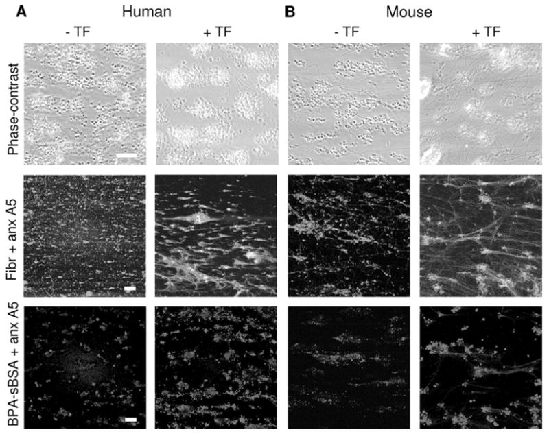
Heterogeneity in human and murine thrombi formed on collagen. Flow experiments were performed with human (A) or murine (B) blood in the absence of tissue factor (−TF) using PPACK-anticoagulated blood (left columns); or with citrate blood that was perfused together with tissue factor (+TF, 2 pM, f.c.) and CaCl2 (2 mmol/L free Ca2+, f.c.) to allow coagulation (right columns). Standard perfusion time was 4 minutes at a shear rate of 1000 s −1. Blood was preincubated with 0.2 mg/mL OG-fibrinogen. Alternatively, preincubation was with 50 μg/mL BPA-sBSA and postlabeling with 1 μg/mL AF532-labeled streptavidin. In both cases, AF647-annexin A5 was also present. Upper panels, Bright-field phase-contrast images after perfusion. Middle panels, TPLSM images of OG-fibrinogen (green) and AF647-annexin A5 (red) fluorescence (different fields of view). Lower panels, TPLSM images of BPA-sBSA (blue) and AF647-annexin A5 (red) staining. Images are representative of 4 to 8 experiments; bars indicate 20 μm.