
Figure 6.
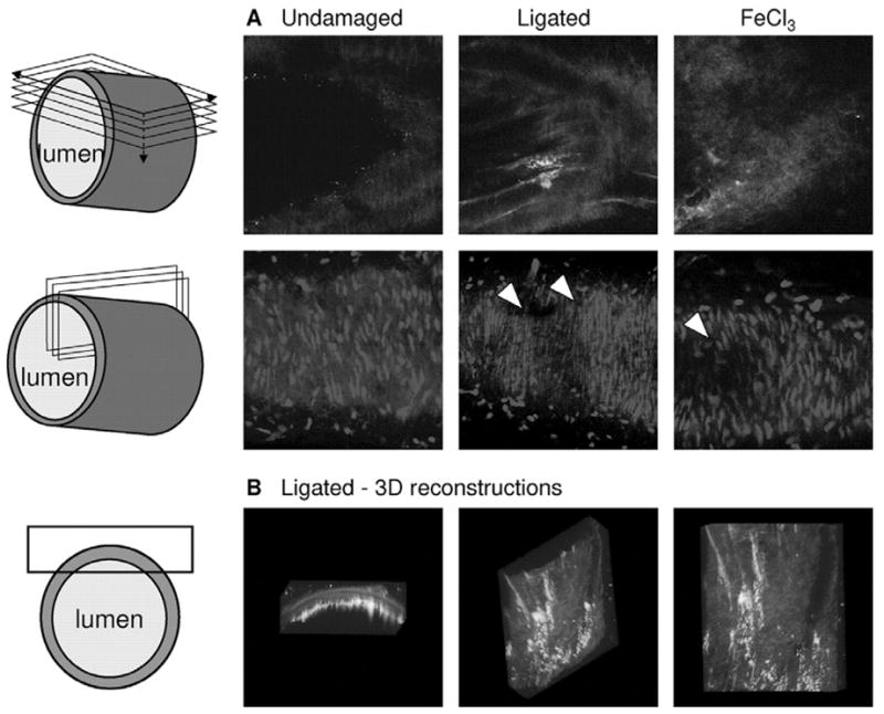
Heterogeneity in arterial thrombi induced by carotid ligation or by FeCl3 application. Mice were infused with OG-fibrinogen and AF568-annexin A5 (200 μg each). A, left panels, Undamaged control carotid artery. Middle panels, One carotid artery was damaged by tight ligation at the bifurcation for 5 minutes to induce vascular damage. Right panels, In other animals, 1 carotid artery was damaged by local application of saturated FeCl3. Thrombus formation proceeded for 10 minutes, after which fluorescence at the luminal side of the artery was recorded by TPLSM. Upper row, Images of fibrin(ogen) (green) and annexin A5 (red) fluorescence at the damaged side of the vessel wall. Note distinct patches of green and red fluorescence; also note blue autofluorescence, indicating the demarcation of the vessel wall with the lumen. Second row, Images of Syto-44 fluorescence (blue) at cross section through the vessel wall. Arrow heads indicate absence of cell nuclei at side of damage. B, Three-dimensional reconstruction of fibrin(ogen) and annexin A5 fluorescence within the ligated artery: side view through vessel wall, turned view, and view from inside of vessel lumen. Data are representative of 3 to 4 vessels. Cartoons indicate way of optical sectioning (images 206 × 206 μm).