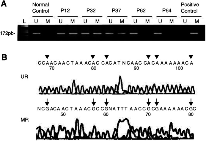
Figure 2.
(A) Methylation-specific PCR of CpG island of the RB1 promoter in glioblastomas (P12 and P32) and anaplastic astrocytoma (P37). Cases P62 and P64 (glioblastomas) showed only unmethylated alleles. Positive control for methylated DNA: normal DNA from lymphocytes treated with SssI methyltransferase; normal control: DNA from a non-neoplastic brain tissue. Negative control from untreated lymphocytes DNA is not shown (L: molecular weight marker). (B) Sequence (reverse of the coding strand) analysis of bisulphite-modified DNA from tumour P12 (MR) and from non-neoplastic brain tissue (UR). Tumour DNA shows methylated cytosines (G in the reverse sequencing marked by arrows) at the represented CpG sites, whereas all CpG cytosines are unmethylated in DNA from non-neoplastic brain tissue (A in the reverse sequencing marked by arrowheads).

