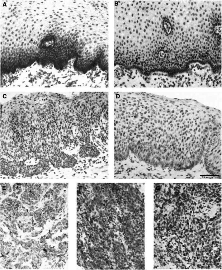
Figure 1.
Immunohistochemistry for detection of survivin in (A) normal oesophageal squamous cell epithelium, (C) high-grade dysplasia (predominant cytoplasmic localisation), (D) high-grade dysplasia (predominant nuclear localisation), (E) oesophageal SCC (cytoplasmic localisation), (F) oesophageal SCC (nuclear localisation). Immunohistochemistry for detection of ki-67 antigen in (B) normal oesophageal squamous cell epithelium (paired section to (A)) and (G) oesophageal SCC (paired section to (F)). Scale bar=100 μm.

