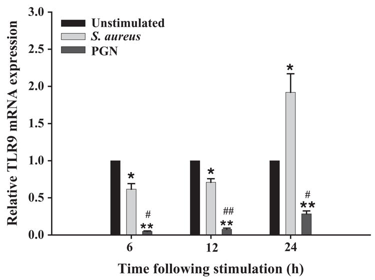
Fig. 8.

S. aureus and PGN differentially modulate TLR9 expression. The time course profile of TLR9 mRNA expression following exposure to either 107 heat-inactivated S. aureus or 10 μg/ml PGN in CD14 WT microglia was measured by qRT-PCR as described in the Materials and methods section. Each real-time PCR reaction was performed in duplicate for TLR9 and the ‘‘housekeeping’’ gene GAPDH. The level of gene expression was calculated after normalizing TLR9 signals against GAPDH and is presented in relative mRNA expression units (mean ± S.D. S.D. of fold-changes pooled from three independent experiments). Significant differences between unstimulated versus bacterially stimulated microglia are denoted with asterisks (*p <0.05; **p <0.001, whereas significant differences between S. aureus- and PGN-treated microglia are also indicated (*p <0.05; **p <0.001).
