
Figure 8.
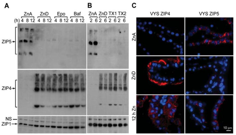
Effects of proteosome and lysosome pathway inhibitors or zinc on ZIP5 and ZIP4 protein abundance and localization in the visceral yolk sac (VYS) explant cultures.
VYS were harvested from d14 fetuses carried in pregnant females that had been fed a zinc-adequate (ZnA) diet or a zinc-deficient (Zn-D) diet beginning on d8 of pregnancy. VYS were then cultured in vitro under the conditions described below. (A) VYS from zinc-adequate dams were incubated in zinc-adequate medium (DMEM containing 10% FBS; ZnA). VYS from zinc-deficient dams were incubated in zinc-deficient medium (DMEM containing 10% Chelex-treated FBS with DMSO vehicle; ZnD), or ZnD medium also containing 50 nM synthetic epoxomicin (Epo) or 100 nM bafilomycin A1 (Baf) for the indicated times (up to 12 h). ZIP5 was immunoprecipitated (top left panel) and detected as described in the legend of Figure 6B. An equal amount of total membrane protein from each sample was examined by Western blot analysis for ZIP4 (middle panels) and ZIP1 as a loading control (bottom panels). (B) VYS from zinc-adequate dams were incubated in ZnA medium. VYS from zinc-deficient dams were incubated in ZnD medium plus or minus a mixture of 1 μM epoxomicin, 50 μM MG132 and 50 μM clasto-lactacystin β-lactone (TX1) or 1 μM epoxomicin and 50 μM clasto-lactacystin β-lactone (TX2) for the indicated times. ZIP5, ZIP4 and ZIP1 detected by Western blot analysis as described in (A). (C) VYS from zinc-adequate dams were incubated for 12 h in ZnA medium, as above. VYS from zinc-deficient dams were incubated for 12 h in ZnD medium ±100 μM ZnCl2 (12 h Zn). VYS were fixed and processed, and ZIP4 and ZIP5 were detected by immunofluorescence as described in the legend of Figure 4. Blue: nuclei; red: sites of antibody binding.