Figure 9.

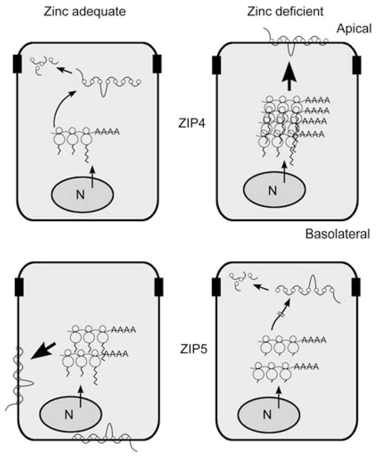
Cartoon summarizing mechanisms by which ZIP4 and ZIP5 are reciprocally regulated by zinc in the same cell. The mouse Zip4 and Zip5 genes are actively expressed in the intestine and fetal visceral yolk sac (VYS). Zip5 is also actively expressed in pancreatic acinar cells. This cartoon depicts the polarized intestinal enterocyte. N, nucleus. Polysomal mRNAs are indicated as lines ending in AAAA and bound by three ribosomes, with the protein tails extending from the ribosomes. ZIP4 and ZIP5 proteins are indicated as lines connected by eight circles representing the eight transmembrane domains. The degraded proteins are indicated as individual circles and lines inside the cells. Zinc apparently does not alter the rate of transcription of either Zip4 (top) or Zip5 (bottom), as indicated by the arrows on the nuclei in each panel. Large bold arrows indicate the most active expression of each of these zinc transporters. Top left: when zinc is adequate, Zip4 mRNA is unstable and ZIP4 protein is not detected on the apical membrane, but is degraded. Top right: when zinc becomes deficient, Zip4 mRNA is stabilized, leading to increased accumulation of Zip4 mRNA and ZIP4 protein and the localization of ZIP4 at the apical membrane. Repletion of zinc causes the rapid endocytosis and ultimate degradation of ZIP4 and destabilization of Zip4 mRNA. Bottom left: Zip5 mRNA abundance remains unchanged in response to zinc. When zinc is adequate, Zip5 mRNA is translated and ZIP5 protein accumulates in the basolateral membrane. Bottom right: when zinc becomes deficient, Zip5 mRNA translation is arrested (hatched arrow), but this mRNA remains associated with polysomes. ZIP5 protein is removed from the membrane and degraded. Repletion of zinc causes the rapid resynthesis and relocalization of this protein to the basolateral membrane.
