
Figure 3.
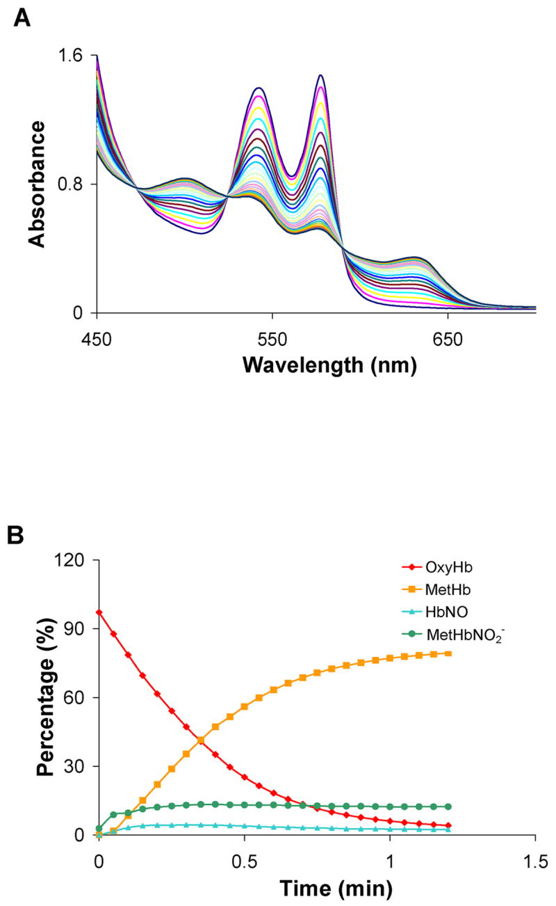
The reaction of Angeli’s salt with a molar excess of oxyHb. OxyHb (100 μM) was mixed with 50 μM Angeli’s salt in 0.1 M phosphate buffer under aerobic conditions. (A) UV-Vis spectra were recorded at 3.0 min intervals after the initial scan. (B) Each spectrum was fit to basis spectra to determine the percentage of each species at each time point. Data from a representative experiment are shown. The average amount of each species formed at 72 minutes from three different experiments was 3 ± 2 % oxyHb, 83 ± 5 % metHb, 2 ± 1 % HbNO, and 10 ± 4 % nitrite bound metHb. The remainder (about 2%) was fit as deoxyHb.