
Figure 5.
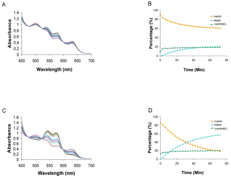
The reaction between metHb with Angeli’s salt. (A) MetHb (100 μM) was mixed with 100 μM Angeli’s salt in 0.1 M phosphate buffer equilibrated in aerobic conditions. Spectra are shown at 3 minute intervals over 72 minutes. (B) Each spectrum was fit to basis spectra to determine the percentage of each species at each time point. After 72 minutes, we found there to be 20% ± 2% HbNO, 64% ± 3% metHb, 15% ± 5% metHb-NO2− (n=3) (C) metHb (100 μM) was mixed with 100 μM Angeli’s salt in 0.1 M phosphate buffer equilibrated in anaerobic conditions. Spectra are shown at 3 minute intervals over 72 minutes. (D) Each spectrum was fit to basis spectra to determine the percentage of each species at each time point. After 72 minutes, we found there to be, 16% ± 9% metHb, 22% ± 3% metHb-NO2−, 54% ± 5% HbNO (n=3).