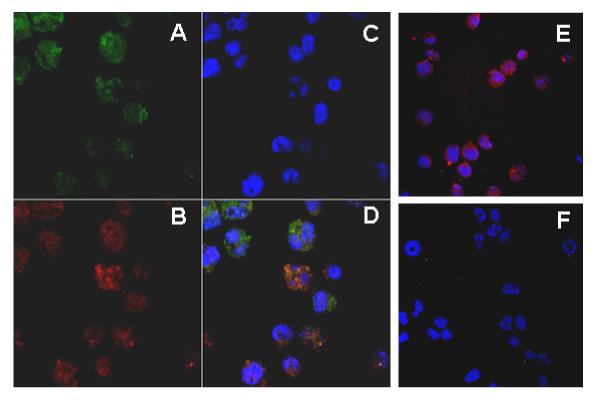
Figure 2. LRG exhibited the same cellular localization as that of myeloperoxidase (MPO).

32Dcl3 cells stably transfected with V5 epitope-tagged mLRG or vector alone were cytospun onto slides and fixed with 4% formaldehyde. LRG and MPO were detected by mouse monoclonal anti-V5 and goat anti-human MPO primary antibodies, which were then detected using TRITC-conjugated donkey anti-mouse IgG and Cy5-conjugated donkey anti-goat IgG secondary antibodies, respectively. Nuclei were stained with Hoescht. (A) Cellular localization of LRG in LRG-transfected cells. (B) Cellular localization of MPO in LRG transfectants. (C) Nuclear staining with Hoescht in LRG transfectants. (D) Merged figure ofA, B and C. (E) Cells transfected with vector alone only stained positively for MPO. (F) LRG transfectants stained with secondary antibodies only.
