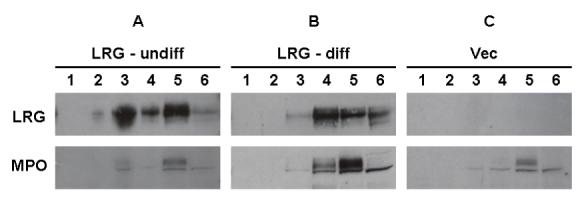
Figure 3. Analysis of subcellular fractions from 32Dwt18 transfectants.

32Dwt18 cells stably transfected with V5-tagged murine LRG (undifferentiated, A; differentiated, B) or with empty vector only (C) were resuspended in disruption buffer, and disrupted by nitrogen cavitation. The nuclei and intact cells were pelleted by centrifugation, and the resultant postnuclear supernatants were loaded onto Percoll gradients. After centrifugation, six continuous fractions were collected. After ultracentrifugation to remove Percoll, the biological materials were collected, and were then subjected toSDS-PAGE analysis. Samples were immunoblotted with an antibody to the V5 epitope (upper panels) or murine MPO (lower panels).
