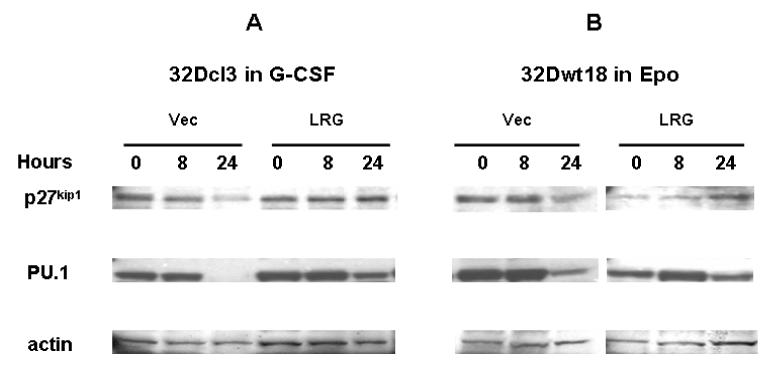
Figure 7. p27kip1 and PU.1 display similar patterns of expression in LRG-transfected 32Dcl3 and 32Dwt18 cells.

Cells were washed out of IL-3 containing media and transferred to media containing G-CSF or Epo as indicated. Whole cell lysates were made from cells harvested at the indicated time points and used for immunoblotting with p27kip1 antibody (top panels). The membrane was stripped and reblotted with PU.1 (middle panels) and actin antibodies (bottom panels). (A) Vector or LRG transfected 32Dcl3 cells in G-CSF containing media. (B) Vector or LRG transfected 32Dwt18 cells in Epo containing media.
