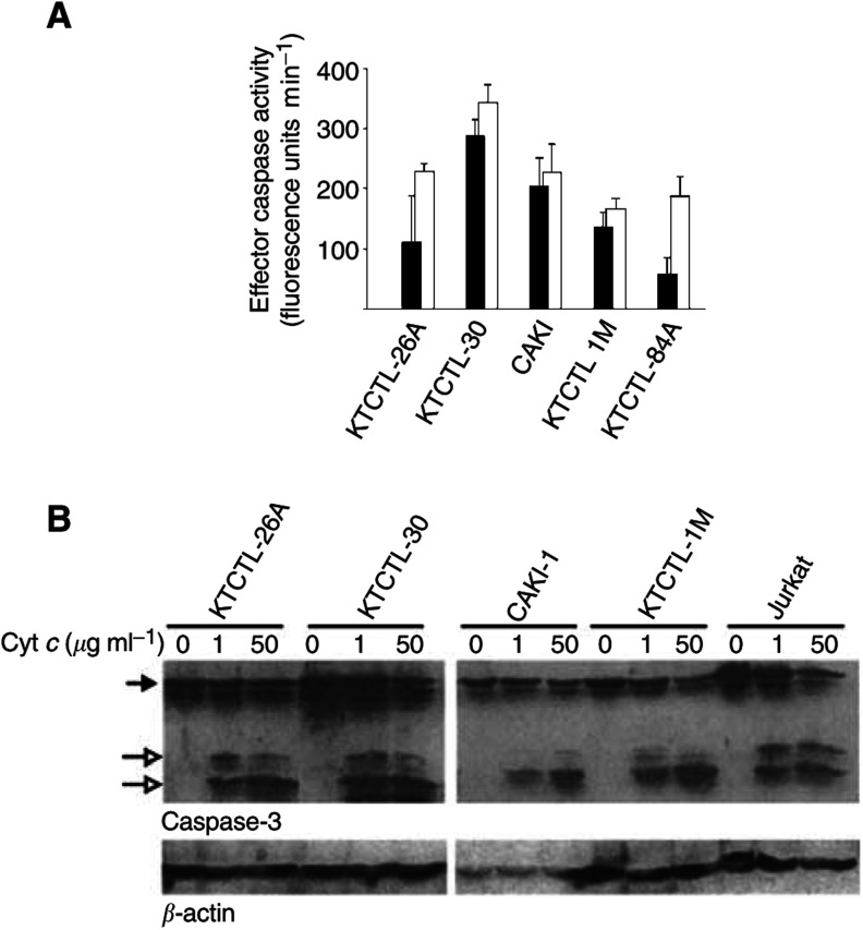
Figure 1.
Composition and function of the apoptosome in five RCC cell lines. (A) Extracts from five RCC cell lines were incubated for 1 h at 37°C in the presence of cytochrome c (one (filled bars) or 50 (open bars) μg ml)−1. Then, DEVD-cleaving activity was measured as release of AMC from the effector caspase-substrate DEVD-AMC (see Materials and Methods for details of the method). Columns give means and s.d. of the results from three separate experiments, each with separately prepared extracts. (B) The presence and activation of caspase-3 in RCC cell line extracts, extracts from the T-cell line Jurkat for comparison. Extracts were incubated with cytochrome c as above, and caspase-3 and β-actin were detected by immunoblotting (200 μg of total protein per lane was loaded). Filled arrow, procaspase-3, open arrows, processed caspase-3. Results for caspase-3, -7 and -9 are summarised in Table 1.

