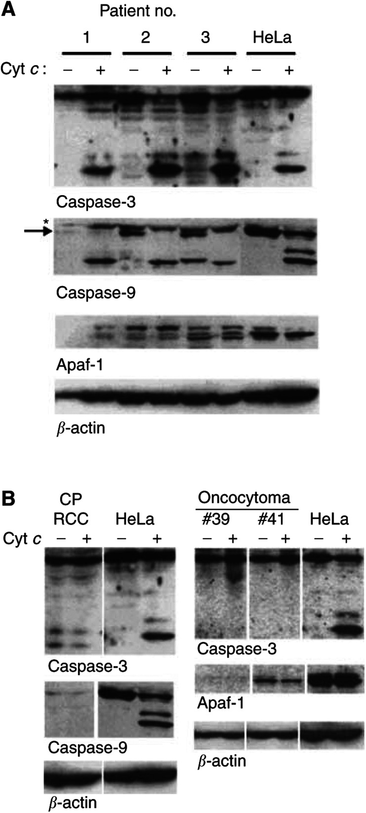
Figure 2.
Examples of expression and processing of components of the apoptosome in clinical samples from RCC. Cells were isolated and extracts were prepared from fresh explants of clear cell RCC (A) or from one chromophobe RCC and two oncocytomas (B). Extracts from HeLa cells were prepared as above. Extracts (800 μg protein in 40 μl) were incubated for 1 h at 37°C in the presence or absence of 50 μg ml−1 cytochrome c. A measure of 200 μg per lane were run on SDS–PAGE and proteins were detected by Western blotting. Purity of the chromophobe RCC was about 80% tumour cells, suggesting that a contamination of 20% nontumour cells does not distort the results. (A) Asterisk denotes a nonspecific band, arrow procaspase-9. The several bands recognised by the anti-Apaf-1 antibody were seen in several experiments and may constitute Apaf-1 variants or, at least in part, products of nonspecific degradation. The smaller size band in the lane patient #3, no cytochrome c in the caspase-9 blot is of unknown origin and was not see in any other blot.

