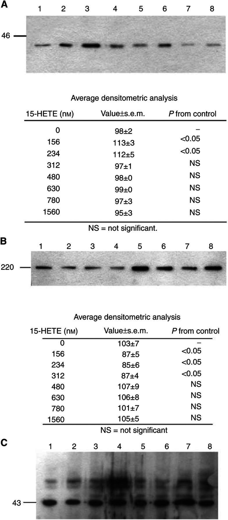
Figure 7.
Western blots of soluble extracts of C2C12 myotubes treated with 0 (lane 1), 156 nm (lane 2), 234 nm (lane 3), 312 nm (lane 4), 480 nm (lane 5), 630 nm (lane 6), 780 nm (lane 7) and 1560 nM 15(S)-HETE, (lane 8) for 24 h on expression of p42 subunit of 19S regulatory subunit (A), myosin (B) and actin control (C). Blots were developed with the respective antibodies as described in Materials and methods, and are representative of three separate determinations on different samples. Average densitometric data are presented under the blots.

