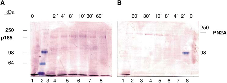
Figure 2.
Western blot analysis demonstrating the phosphorylation state specifity of the PN2A antibody. Specifity of (A) anti-Her-2/neu (p185) antibody and (B) anti-phospho (tyr1248) Her-2/neu antibody (PN2A) is shown by Western blot analysis of whole-cell lysates. SKBR3 cells were treated without (lanes A1 and B8) and with 100 ng ml−1 EGF for 2 (lanes A3 and B6), 4 (lanes A4 and B5), 8 (lanes A5 and B4), 10 (lanes A6 and B3), 30 (lanes A7 and B2) and 60 (lanes A8 and B1) min (kDa, molecular weight in kilodalton, molecular weight markers at 250, 98 and 64 kDa).

