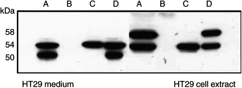
Figure 5.
Recovery of CA IX bands 50 and 58 kDa from diluted fluids after immunoprecipitation. A=original TC medium or extract from HT29 cells. To the same antigens as in A was added mAb M75 (ascites fluid) and Protein A-Sepharose and after appropriate incubation, the mixtures were centrifuged and analysed; B=supernatant; C=pellet. With antigens from the medium, B+C was mixed and analysed=D; with antigen from cell extract, to the pellet was added 2% FCS (=D). All samples for the analysis were adjusted to the same concentration of CA IX protein.

