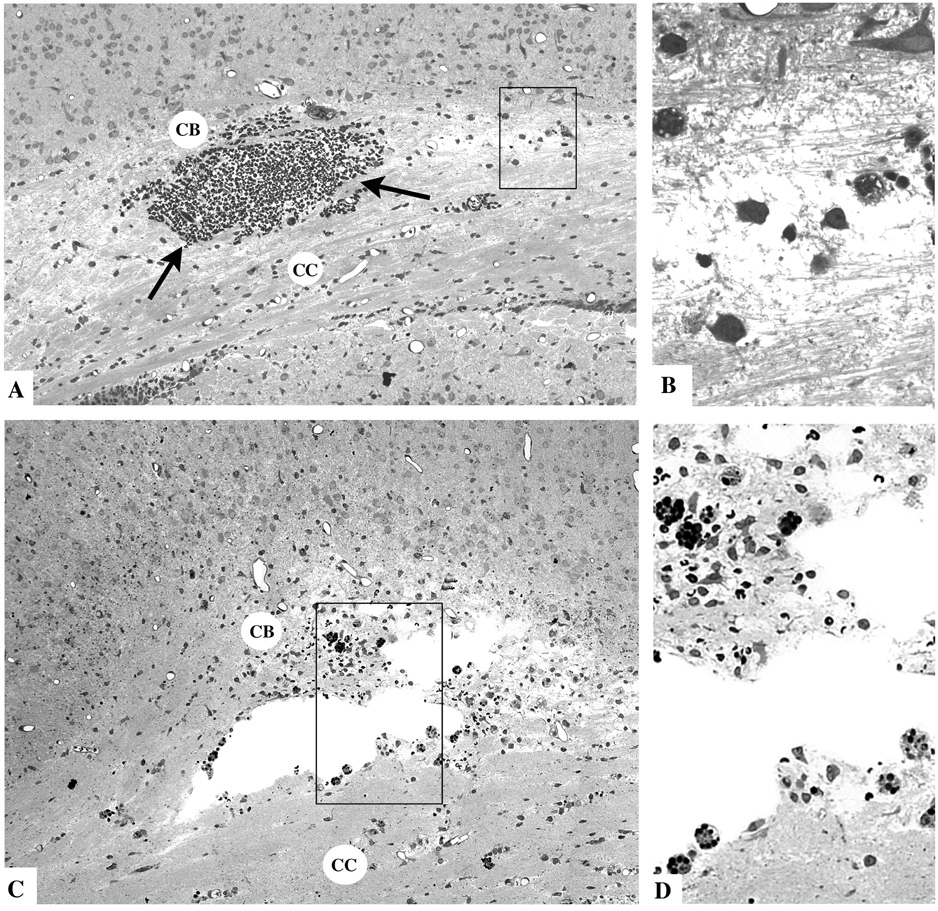
Fig. 2.
Thin plastic sections from the corpus callosum (CC) and cingulum bundle (CB) region of the infant rat brain 4 hours (A and B) or 16 hours (C and D) after TBI. The typical findings at 4 hours are a mild degree of interstitial edema giving portions of the CCCB track a rarefied appearance, and focal accumulations of red blood cells (arrows) due apparently to rupture of small blood vessels. The boxed region in A, shown at higher magnification in B, exhibits axonal fibers in disarray, surrounded by edema fluid and accompanied by phagocytic cells that are just beginning to respond to the pathological reaction. At 16 hrs, cystic spaces have formed at the site of tissue injury. The boxed regions in C, shown at higher magnification in D, reveals many phagocytic cells lining the cystic spaces and occupying the surrounding tissue area where they are ingesting red blood cells (dark round masses) and other less dense debris.

