
Fig. 6.
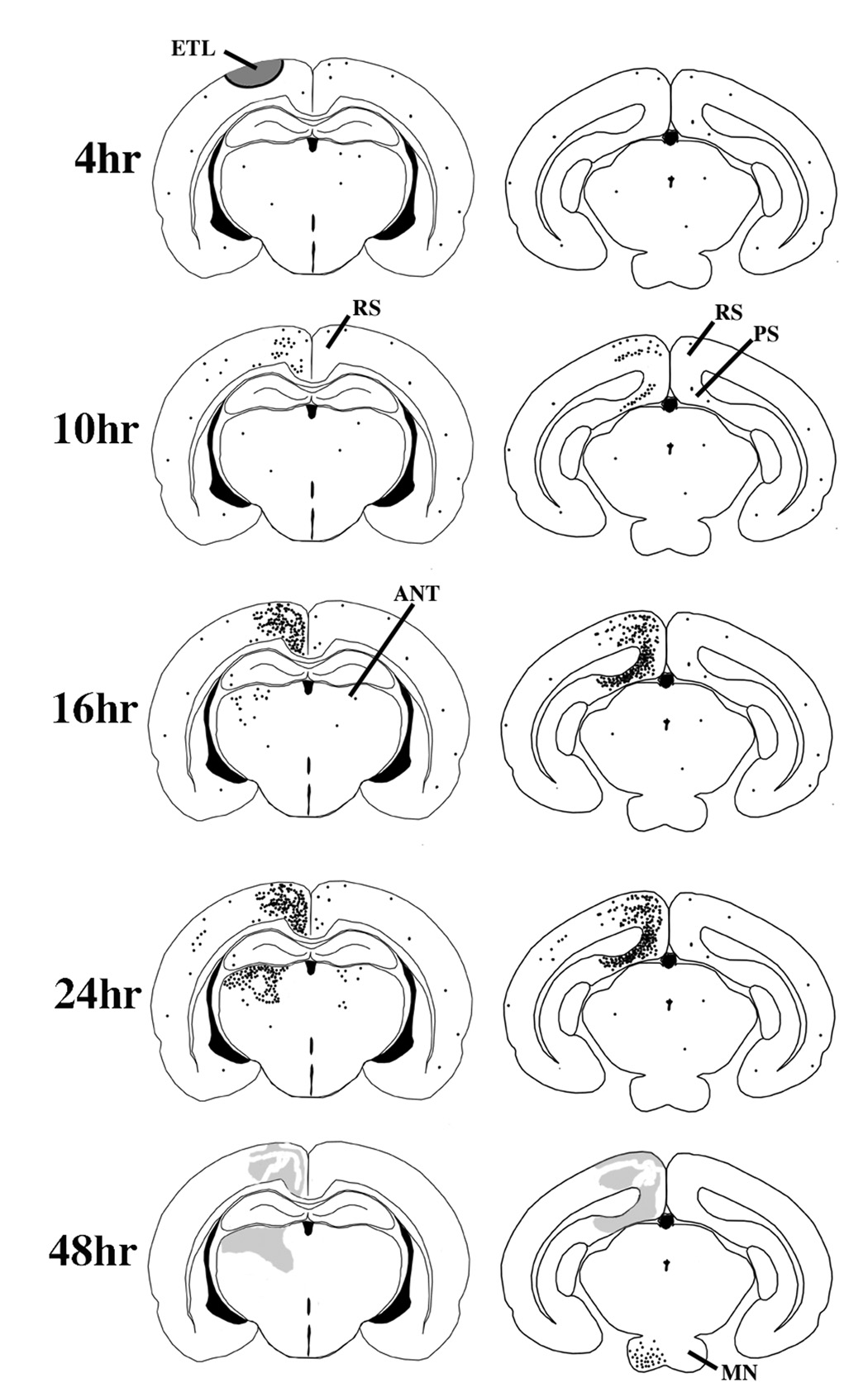
This schematic summarizes the nature, time course and localization of pathological changes in the brain following relatively mild concussive impact to the head of a P7 rat. At 4 hrs post-impact, AC-3 staining does not reveal any pathological reaction, but other histological procedures demonstrate an acute excitotoxic lesion (ETL) that rapidly kills neurons in the semicircular zone shaded in grey. At 8 – 10 hrs AC-3 staining begins to reveal a wave of neuroapoptosis affecting the ipsilateral retrosplenial (RS) and pre/para-subicular (PS) cortices. By 16 hrs, the RS and PS cortical involvement has progressed substantially, and a new wave of neuroapoptosis begins to appear in the ipsilateral anterior thalmic nuclei (ANT). By 24 hrs the ANT lesion has progressed to its full extent and there is mild increased staining in the contralateral ANT and RS and also in scattered neurons in the ipsilateral cortical mantle. In the 36–48 hr interval a third wave of neuroapoptosis appears in the ipsilateral mammillary nuclei (MN) and the apoptotic lesions in other brain regions are no longer visible, except as vague smudged areas in AC-3 stained sections.