
Figure 7. CRACM1-/- mice have normal T cell development and proliferation but cytokine secretion is inhibited.
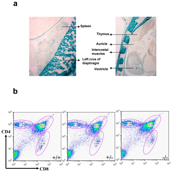

(a) High resolution images of LacZ stained thymus and spleen of 1 day old CRACM1-/- neonates showing absence of staining in thymic lymphoid areas. Staining was done as described in Figure 1d. (b) Thymus staining for CD4 and CD8 antigens. Thymocytes harvested from 5 week old CRACM1 +/+ (left), +/- (middle) and -/- (right) littermates were stained with CD4-PE-Cy5 and CD8-FITC and analyzed using flowcytometry (c) Proliferation of CRACM1 +/+ (diamonds), +/- (squares) and -/- (triangles) splenocytes. Splenocytes were stimulated with different doses of anti-CD3 + anti-CD28 antibodies for 48 hrs and pulsed with 3H-thymidine for the last 6 hrs. Results are mean ± SEM of triplicates, representative of n=4. (d) IL-2 and (e) IFN-γ secretion by CRACM1 +/+ (diamonds), +/- (squares) and -/- (triangles) splenocytes stimulated as described in Fig. 2c. 48 hrs post-stimulation supernatants were collected and cytokine levels estimated. Results are from pooled triplicates, representative of n=4. (f) Real time PCR for CRACM1, CRACM2 and CRACM3 mRNA in thymocytes isolated from CRACM1 +/+ (hashed bars), +/- (dotted bars) and -/- (open bars) littermates.