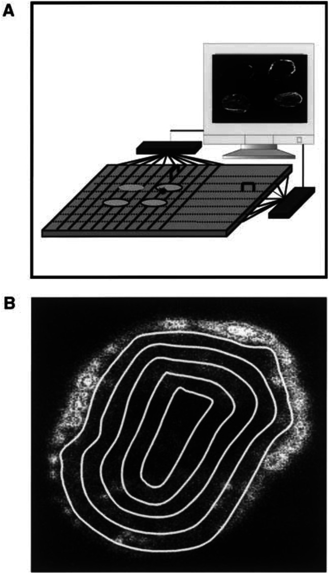
Figure 1.
(A) Schematic illustration of the Bioscope system. The sensor of the system is a 300-μm-thick double-sided silicon strip detector with a sensitive area of 32×32 mm. The detector has 640 strips in each direction. Four tumour slices can be imaged simultaneously. (B) Bioscope image of an A-07 tumour. The concentration of 99mTc in the tumour tissue is proportional to the intensity in the image. Quantitative analysis of the radial distribution of 99mTc was performed by dividing the images into five sectors and analysing each sector separately. The sectors were bounded by lines drawn at distances of nR/5 from the tumour centre, where R is the tumour radius and n is the sector number.

