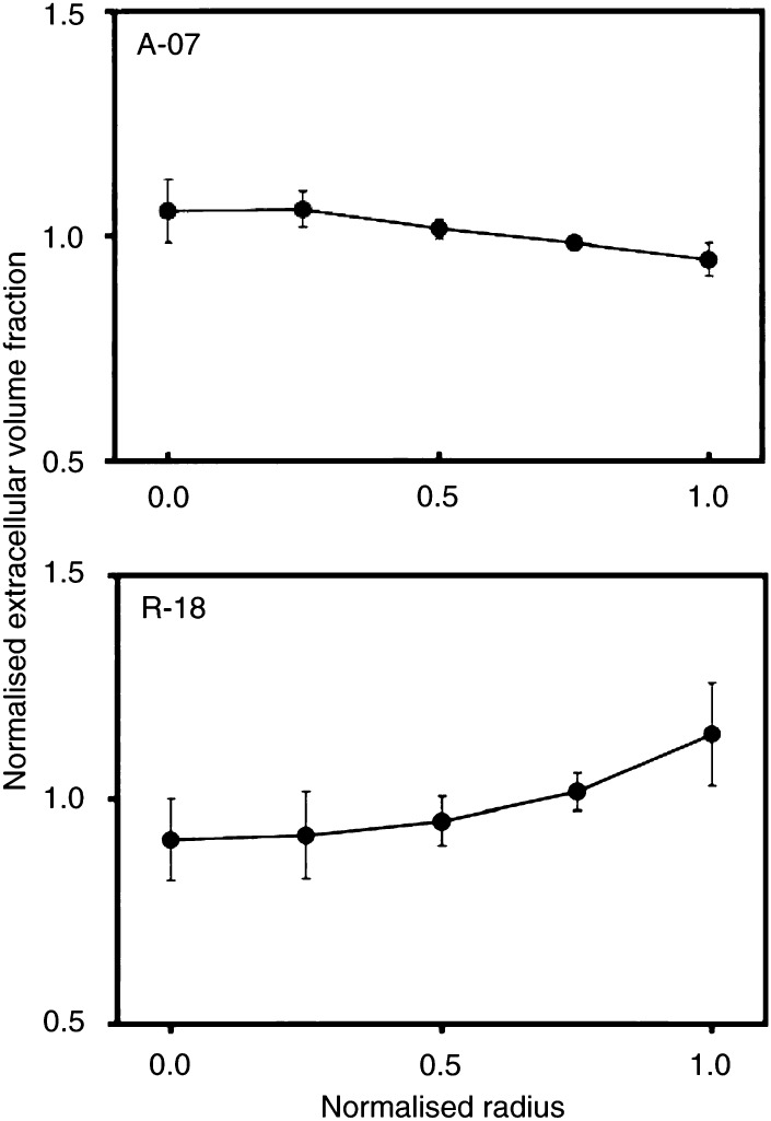
Figure 7.
Extracellular volume fraction vs distance from tumour centre in A-07 and R-18 tumours. 99mTc-HSA was administered intravenously to tumour-bearing mice with ligated renal arteries. The mice were killed and the tumours were frozen in liquid nitrogen 3 h after the administration of 99mTc-HSA. The concentration of 99mTc-HSA in the tumour tissue was used as a parameter for extracellular volume fraction. Tumour size was normalised, that is, tumour radius was assigned a value of 1.0. Extracellular volume fraction was also normalised, that is, the mean concentration of 99mTc-HSA in each tumour section was assigned a value of 1.0. One section was analysed for each tumour. Points represent mean values of 8 (A-07) or 7 (R-18) tumours. Bars represent s.e.m.

