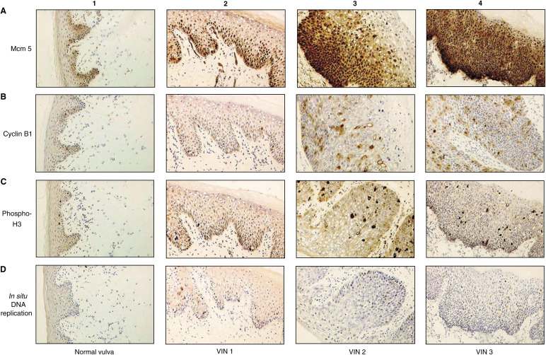
Figure 1.
Immunostaining of normal vulval epithelium and VIN 1, VIN 2 and VIN 3 with cell cycle markers (x40): (A1) normal vulval epithelium shows that Mcm 5 expression is restricted to the basal epithelial layer; (A2) in VIN 1, >40% of cells express Mcm 5 in the basal and suprabasal epithelial layers; (A3) in VIN 2, >85% of cells express Mcm 5 and positive nuclei are detected in basal and superficial epithelial layers; (A4) VIN 3 shows >90% of cells express Mcm 5, extending throughout the full thickness of the epithelium; (B1) in normal vulval epithelium, expression of cyclin; (B1) is restricted to the basal epithelial layer. In VIN 1 (B2), VIN 2 (B3) and VIN 3 (B4), a greater proportion of cells express cyclin B1 compared with normal skin and positively stained nuclei are found in progressively more superficial epithelial layers, correlating with the level of dysplasia. C1–4 and D1–4 show a similar pattern of expression of phosphohistone H3 in normal vulva (C1), VIN 1 (C2), VIN 2 (C3) and VIN 3 (C4), and by in situ DNA replication in normal vulva (D1), VIN 1 (D2), VIN 2 (D3) and VIN 3 (D4).

