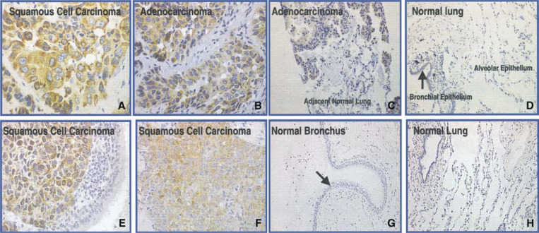
Figure 4.
Immunohistochemistry analysis of L523S using affinity-purified whole IgG molecules (A–D) and F(ab) fragments (E–H). Illustrated here are results of IHC staining on tissue sections of LSCC (A, E, F), lung adenocarcinoma (B), lung adenocarcinoma with adjacent normal lung tissue (C), and normal tissues of lung (D, H). Homogeneous cytoplasm staining was observed in both squamous and adenocarcinoma samples (A–C, E, F). Arrows point to bronchiole epithelial cells that were lightly stained with whole IgG molecules (D), but not with F(ab) fragments (G).

