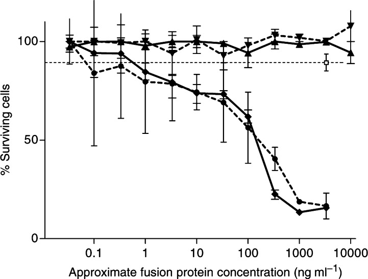
Figure 2.
A33scFv–CD-mediated cytotoxicity on A33 antigen-positive vs negative cells: LIM1215 cells and HT29 cells were incubated with a dilution series of A33scFv–CD fusion protein and, after washing, with the 5-FC prodrug. Survival was measured by the MTT method as described. A33scFv–CD fusion protein from two different preparations was used on HT29 cells (▴ and ▾) and on LIM1215 cells (• and ⧫). As a control, a single, high concentration of A33scFv–GFP (□) was used instead of A33scFv–CD. Mean and s.d. of triplicate samples.

