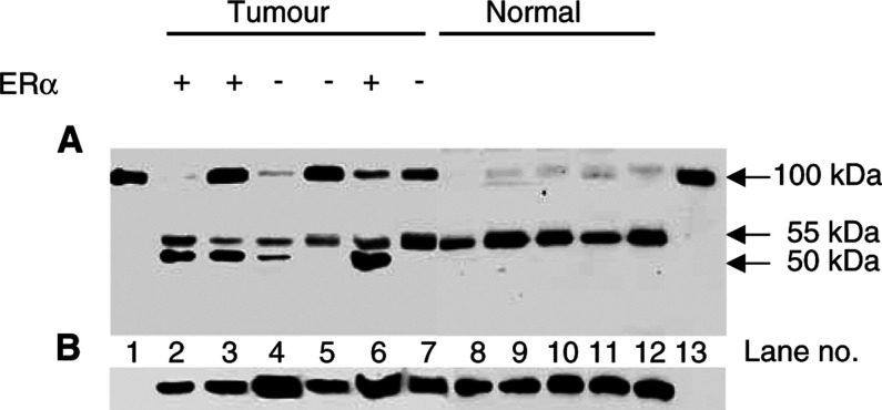
Figure 2.
Analysis of fibulin-1 protein expression in representative breast tissue extracts. In (A), purified human fibulin-1 protein (lanes 1 and 13) and 40 μg of breast tumour extracts (lanes 2–7) and 40 μg of normal breast tissue (lanes 8–12) extract. Samples were run on a 10% polyacrylamide gel under reducing conditions, transferred to PVDF membrane and probed with the 3A11 mAb, which is directed against the N-terminus of human fibulin-1. In addition to the mature fibulin-1 polypeptide (apparent molecular mass of 100 kDa) immunoreactive fragments of 55 and 50 kDa are detectable. The ER status of the breast tumours is also indicated. In (B), the blot shown in (A) was stripped and reprobed with a panspecific antiactin antibody to serve as a control for protein loading.

