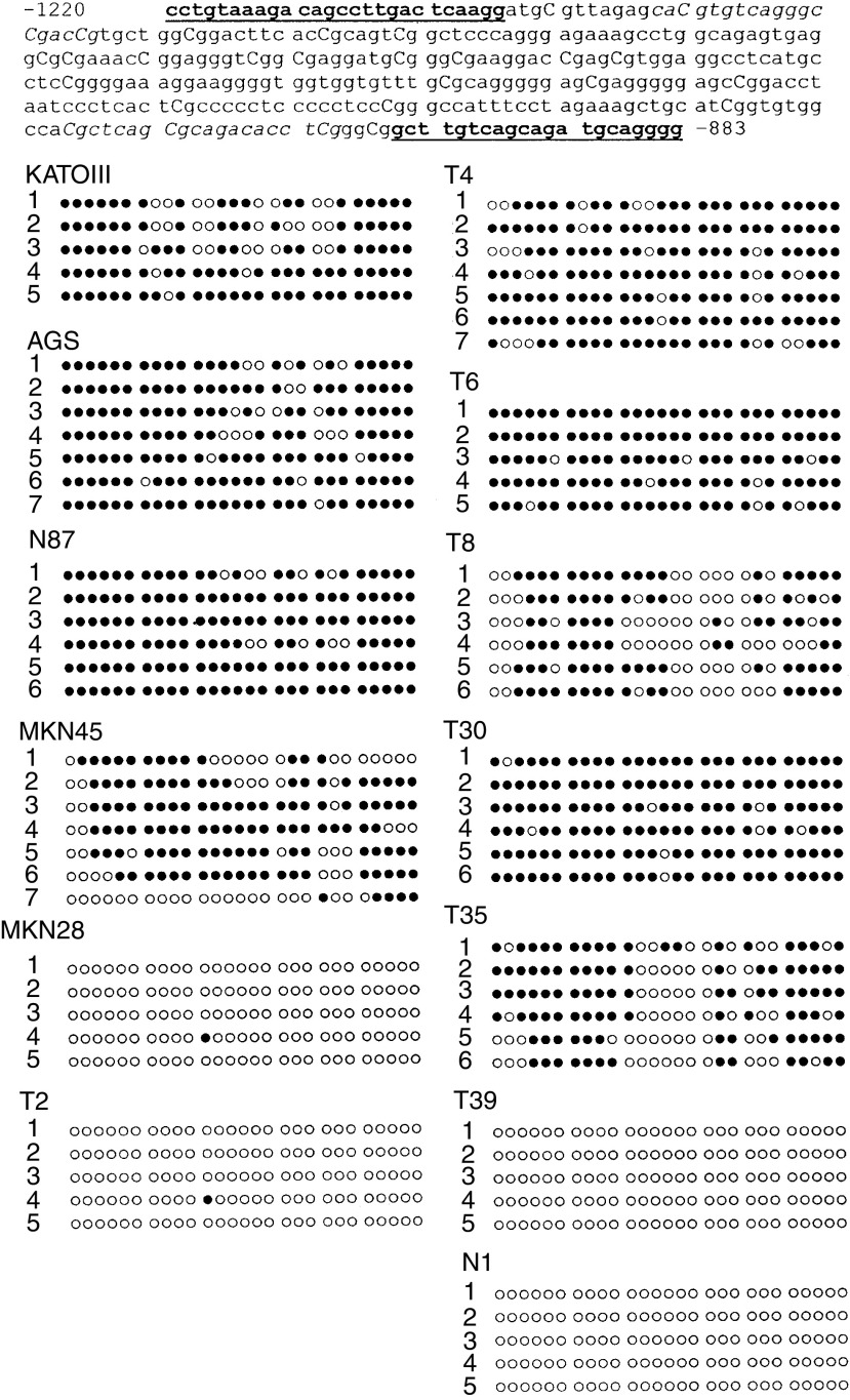
Figure 4.
Bisulphite sequencing of cyclin D2 promoter region. The nucleotide sequence from −1220 to −883 of the cyclin D2 gene is shown. The individual CpG sites between two PCR primers are numbered sequentially. Cytosines at the CpG site are in capitalisation. The bisulphite sequencing PCR primers are bold and underlined whereas the MSP primers are shown as italic. DNA from five gastric cell lines, two cyclin D2-positive cancers (T2, T39), two cancers with low cyclin D2 expression (T8, T39) and three cyclin D2-negative (T4, T6, T30) cancers as well as one normal gastric tissue (N1) were bisulphite-treated, PCR-amplified and subcloned. The sequencing results from five to eight clones for each cell line and samples are presented. Each horizontal line represents the sequencing result of one subclone. CpG sites within 48 bp are shown as one block. Methylated CpG sites are shown as ‘•’ whereas ‘○’ indicate unmethylated CpG sites.

