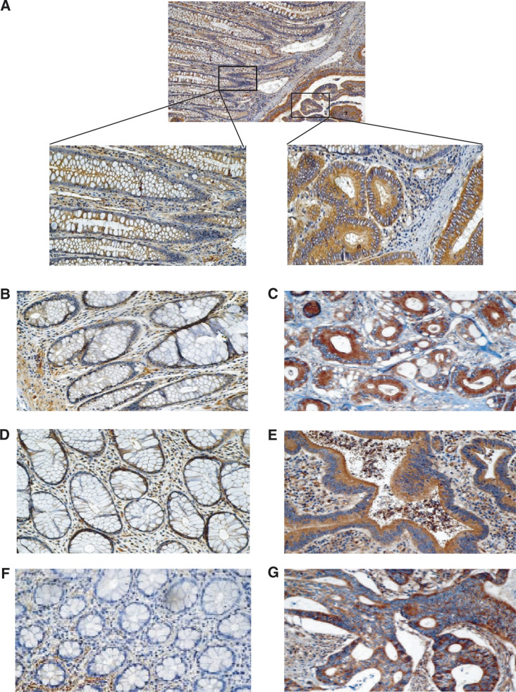
Figure 1.
Over-expression of ILK in sporadic colorectal cancers. Panel A: representative case examining ILK expression in the control crypts vs cancerous crypts at a lower magnification (× 100) as well as at a higher magnification (× 200). Panels B–G: three additional representative cases demonstrating enhanced ILK expression in the cancerous lesions (C, E, G) when compared with the normal control (B, D, F). Staining was performed as outlined in the Materials and Methods section.

