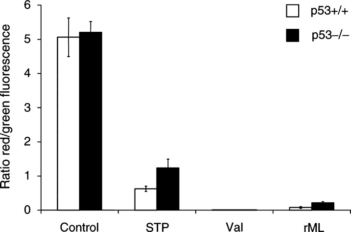
Figure 3.
Loss of mitochondrial membrane potential by rML. The mitochondrial membrane potential of p53-wild-type and p53−/− deficient E1A/ras-transformed MEFs was analysed with JC-1 6 h after rML-treatment (0.5 ng ml−1). Control experiments were performed with valinomycin (50 nM, 15 min) and staurosporine (1 μM, 3 h). The alterations from JC-1 aggregates (red fluorescence) to JC-1 monomer (green fluorescence) are presented as mean ratio (red/green fluorescence)+s.d. from two independent experiments.

