Figure 3.
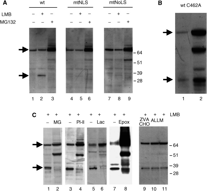
The appearance of the 32 kDa is a post-transcriptional event and is prevented by proteasome inhibitors. (A) H1299 cells were transfected with vectors expressing either wild-type hMdm2 lanes (1–3), hMdm2 mtNLS lanes (4–6) or hMdm2mtNoLS lanes (7–9). In lanes 1, 4 and 7, cells were left untreated. In lanes 2, 5 and 8, they were treated with LMB, in lanes 3, 6 and 9, they were treated for 3 h with the proteasome inhibitor MG132 (20 μM). Cells were harvested and hMdm2 was detected by Western blotting as above. The positions of the bands corresponding to the full-length hMdm2 and the 32 kDa fragment are indicated by arrows. (B) H1299 cells were transfected with an expression vector for hMdm2 or the hMdm2C462A mutant and treated with 2 nM leptomycin B for 18 h. hMdm2 was detected as above. (C) H1299 cells were transfected with 5 μg of the expression vector for hMdm2 (pCMVhMdm2) and treated with 2 nM leptomycin B alone for 18 h (lanes 1, 3, 5, 7 and 11) or together with 20 μM MG132 (lane 2), 20 μM PI-II (lane 4) or 20 μM lactacystin (lane 6), 4 μM epoxomycin (lane 8), 10 μM Z-Val-Phe-CHO (lane 9) or 10 μM ALLM (lane 10). Cell extracts were analysed by Western blotting and hMdm2 was detected using 4B2. The positions of the bands corresponding to the full-length hMdm2 and the 32 kDa fragment are indicated by arrows on the left side.

