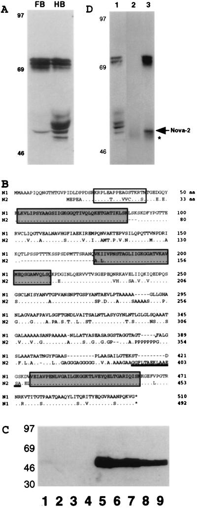
Figure 1.
Identification of Nova-2, a PND antigen. (A) Western blot analysis of POMA antigens in P0 mouse brain. Total protein extracts (50 μg per lane) from P0 mouse forebrain (FB) or hindbrain (HB) were analyzed by Western blot analysis with POMA antiserum. Molecular mass markers are indicated on the left (kDa). (B) The full-length human Nova-2 amino acid sequence is shown compared with Nova-1. The Nova-1 sequence includes mini-exon LSK (aa 88–90) and alternatively spliced Exon H (aa 153–176). KH domains are boxed in gray. A potential NLS is boxed in white. The peptide sequence used to generate the Nova-2-specific antibody N2Ab (aa 392–405) is underlined. Dashes indicate gaps inserted to align the sequences, dots indicate identity, and asterisks indicate predicted stop codons. (C) The N2FP is recognized by 5 of 5 POMA patient antisera. Western blots with recombinant N2FP (500 ng per strip) were immunoblotted with POMA antisera (lanes 5–9, serum from five different POMA patients at 1:200, 1:250, 1:250, 1:1000, 1:2000), non-POMA paraneoplastic antisera (lane 3, Yo patient serum at 1:200; lane 4, Nb patient serum at 1:200), or normal human control sera (lane 1, 1:50; lane 2, 1:200). Molecular mass markers are indicated on the left (kDa). (D) The antipeptide antibody N2Ab immunoprecipitates a subset of Nova proteins from mouse brain. Immunoprecipitations from E17 mouse brain extracts were performed with affinity-purified N2Ab (lane 3) or a control affinity-purified rabbit antibody (lane 2). Immunoprecipitates were analyzed for Nova proteins by immunoblotting with POMA antiserum. Extract (50 μg) was loaded (lane 1) to demonstrate the Nova proteins in the starting material. Molecular mass markers are indicated on the left (kDa); the Nova-2 50-kDa band (arrowhead) and IgG bands (asterisk) are indicated on the right.

