Figure 2.
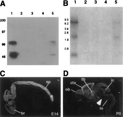
Expression analysis of Nova-2 in mouse. (A) Nova tissue Western blot. Total protein (50 μg per lane) from adult mouse tissues was immunoblotted with POMA antiserum. Lanes: 1, brain; 2, kidney; 3, heart; 4, spleen; 5, lung. Molecular mass markers are indicated on the left (kDa). (B) Nova-2 tissue Northern blot. Total RNA (20 μg per lane) was isolated from adult mouse tissues and hybridized on a Northern blot with a Nova-2 specific probe. Lanes: 1, brain; 2, kidney; 3, heart; 4, spleen; 5, lung. Molecular weight markers are indicated on the left (kb). The blot was stripped and reprobed for actin to demonstrate RNA integrity (data not shown). (C and D) Nova-2 transcripts in the mouse embryo and perinatal mouse. Sagittal sections (12 μm) of E14 embryo (C) or P0 (D) mouse were hybridized with a 33P-radiolabeled Nova-2 riboprobe and imaged with darkfield microscopy (positive signal is white). At E14, Nova-2 mRNA is detected throughout the CNS, including brain (br) and spinal cord (sp). At P0, Nova-2 transcripts are most abundant in the cortex (ctx), with strong signal also in olfactory bulb (ob), thalamus (th), inferior colliculus (ic), and inferior olive (io). Signal is relatively weak in the brainstem (arrowhead). No signal was seen with control sense riboprobes (data not shown).

