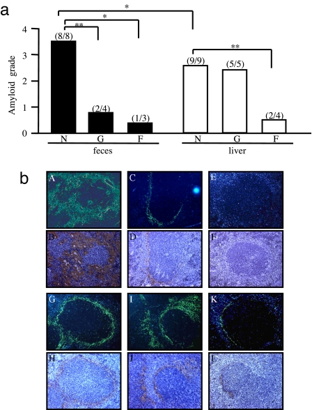
Fig. 3.
High transmissibility of feces from the cheetah with amyloidosis. (a) Fecal (C68 and C67) and liver (C68) AA amyloid fibril fractions were untreated (N), or treated by guanidine-hydrochloride (G) or formic acid (F) and injected into mice to induce AA amyloidosis. Equal quantities of amyloid fractions (100 μg) were used in each experiment. The degree of amyloidosis was determined by the amyloid deposition observed in Congo red-stained sections of the spleen (*, P < 0.05; **, P < 0.01). (b) AA deposition in the spleens of mice exposed to untreated fecal fibrils (A and B) (grade 4) or untreated liver fibrils (G and H) (grade 3). Deposition observed in mice exposed to guanidine-hydrochloride treated fecal fibrils (C and D) (grade 1) or liver fibrils (I and J) (grade 3) and to formic acid treated fecal fibrils (E and F) (grade 0) or liver fibrils (K and L) (grade 1). Deposition was detected by green birefringence in Congo red-stained sections under polarized microscopy (Upper) and immunohistochemical staining with anti-mouse AA antiserum (Lower). (Magnification, × 200.)

