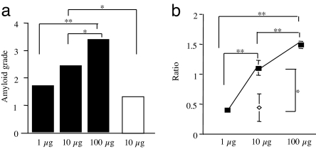
Fig. 4.
Quantitation of transmissibility of AA amyloid fibrils from feces. (a) The degree of amyloid deposition in spleens of mice induced by fecal amyloid fibril fractions (filled squares) (1 μg, 10 μg, and 100 μg) or the liver fractions (open squares) (10 μg) was determined in Congo red-stained tissue sections (four mice per group). (b) The degree of AA deposition in induced mice was determined by isolation of AA amyloid fibril fractions from the spleens of mice in each group (filled squares, fecal; open diamonds, liver) followed by Western blot analysis (20 μg of protein per well) and quantification using National Institutes of Health Images. The means and SE were determined by the relative ratios of AA amyloid protein levels in each group versus the group receiving 10 μg of amyloid fibrils fraction from the feces (*, P < 0.05; **, P < 0.01).

