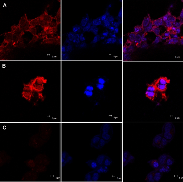
Figure 6. Confocal microscopic images of HEK 293 cells, treated with QD-TRF conjugate (A, B), MSA-CdTe QDs (C).
(B) QDs stained the cells during cell division period. The panel to the left displays the images of CdTe QDs and their corresponding images of HEK 293 cells nuclei stained with Hoechst 33342 are shown in the middle panel. The panel to the right shows the overlays of the above two panels. Laser confocal microscopy images were obtained with laser excitation at 405 nm with a 40×objective.

