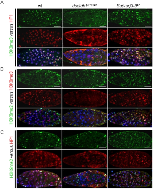
Figure 4. Relationship among di- and tri-methylated H3-K9 patterns, and HP1 pattern in dsetdb1G19561 and Su(var)3–917 germaria.
(A) H3K9me3 versus HP1 patterns. (B) H3K9me2 versus H3K9me3 patterns. (C) H3K9me2 versus HP1 patterns in wild-type (left panel), dsetdb1G19561 (middle panel), and Su(var)3–917 (right panel) flies. Note that HP1 is absent from the inner germarium of the dsetdb1G19561 mutant, from which H3K9me3 signals are absent (in A), whereas H3K9me2 signals remain unaltered (in C). Nuclei were counterstained with 4′,6-diamidino-2-phenylindole (DAPI; blue). Scale bars, 10 µm.

